
The sometimes fuzzy view of microscopic fossils just gained more focus. Using existing digital technology, paleobiologists for the first time viewed intricate structures of tiny fossils in 3-D - a technique that they say could one day aid in the search for life on Mars.
 Compared
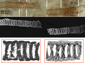
to traditional optical images (top), images in 3-D (middle) can improve the
resolution needed for researchers to see spiral detail in this 650-million-year-old
microscopic fossil from Kazakhstan. Close-up views reveal morphological detail
in the 3-D image (bottom left) as well as the chemical structure imaged with
another device (bottom right), both of which might one day be used to look for
signs of tiny fossils within martian rocks. Image courtesy of William Schopf/UCLA.
Compared
to traditional optical images (top), images in 3-D (middle) can improve the
resolution needed for researchers to see spiral detail in this 650-million-year-old
microscopic fossil from Kazakhstan. Close-up views reveal morphological detail
in the 3-D image (bottom left) as well as the chemical structure imaged with
another device (bottom right), both of which might one day be used to look for
signs of tiny fossils within martian rocks. Image courtesy of William Schopf/UCLA.
William Schopf, a paleobiologist at the University of California in Los Angeles, and colleagues say that 3-D images, captured with a device called confocal laser scanning microscopy (CLSM), offer a simple alternative to slicing and dicing samples. The device also obtains intricate images that can be viewed from any angle. "This is the first technique that permits that," Schopf says.
Previously, researchers viewed tiny fossils by slicing thin sections from a rock, imaging the surface with an optical microscope and then pasting images together into composite sketches. That process, however, depends on the skill of the researcher to reconstruct in their mind how the specimen looks. According to Kathleen Grey, a paleontologist at the Geological Survey of Western Australia in Perth, different interpretations often spark disputes among specialists.
Schopf and colleagues describe CLSM in the January Astrobiology through details they observed on seven different microfossils from Kazakhstan and Australia, dated between 650 million and 850 million years old. The team combined 3-D images of fossil morphology from CLSM with data from an existing instrument that images chemical structure.
Analyses of the resulting 3-D views allowed the researchers to measure the thickness of the fossils' cell walls, discover irregular patterns in the cells and find material that was altogether missing - information that prior technologies could not resolve, Schopf says. The chemical and structural information allows paleobiologists to determine if the process of fossilization has chemically altered a specimen, and also to compare the fossil with modern organisms.
So far, Grey says that she is "impressed" by Schopf's images. One image of a spiral-shaped cyanobacteria struck her because it was similar (the same genus) to a specimen that she had illustrated by compiling individual photographs. The spirals consist of long chains of cells enclosed within a sheath, but such material generally is not well-preserved, Grey says, leaving its identity open to dispute.
The interpretive nature of her illustration, which appeared in both Grey's graduate thesis and a published monograph, caused reviewers of those works to call the structure "not convincingly spiral." But a 3-D image "largely overcomes this problem," Grey says.
Beyond imaging microfossils on Earth, the device could enable researchers to look for microfossils in rocks from Mars. A miniaturized version of the technology could be a component of future unmanned exploration robots on Mars, so that researchers on Earth can look for signs of microfossils within remotely collected images. "It's going to be difficult, but that's possible," Schopf says. Conversely, researchers could look for signs of fossil life inside rock samples returned from a future mission to Mars.
Only 10 years ago, NASA officials thought they found that sign. Daniel Goldin, NASA's administrator at that time, announced on Aug. 6, 1996, that scientists had "compelling" evidence for microscopic life contained within a martian meteorite recovered from Antarctica. But many scientists, including Schopf, remained skeptical and held that the presence of bacteria-like features could be explained by inorganic processes.
The problem was that the necessary tools to look in detail at the chemistry of "objects that they thought might be fossils" were not available, Schopf says. Now, if a similar debate were to arise, "one of the first things you do," he says, is look at its structure in 3-D.
Should researchers find a fossil-like structure on Mars, Grey says that the "high level of proof" to show that it resembles fossils on Earth makes the field of microfossil interpretation "very contentious." Any advance that helps researchers to better interpret tiny fossils, she says, "is welcome."
Kathryn Hansen
 |
Geotimes Home | AGI Home | Information Services | Geoscience Education | Public Policy | Programs | Publications | Careers |