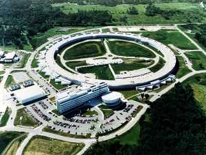
Advanced Photon Source at the Argonne National Laboratory
Courtesy of Neil Sturchio
| Geotimes Home | Calendar | Classifieds | Subscribe | Advertise |
| Geotimes
Published by the American Geological Institute |
November
2000
Newsmagazine of the Earth Sciences |
| INSIDE
ROCKS
By Robert Astheimer,
Kristin Bennett,
|
The tools of physicists are also extraordinary
tools for earth scientists. The research facilities supported by the U.S.
Department of Energys Office of Science provide an unparalleled opportunity
for totally new approaches to understanding earth processes and the properties
of earth materials based on their smallest scale structures. Facilities
include extremely intense, synchrotron x-ray facilities for seeing atomic
structures with electrons and high-powered neutron sources for seeing
atomic structures with neutrons. They provide researchers access to a powerful
array of probes for studying all types of earth materials at the nanometer
to micrometer scales. We can build our knowledge of minerals and fluids
from the inside out, starting with their atoms and working up.
Synchrotron radiation
X-rays are widely used for identifying minerals and determining their
chemical compositions. Historically, the sensitivity of x-ray methods was
limited by the relatively low flux of x-rays produced by conventional sealed-tube
sources. This situation changed dramatically when the first synchrotron
radiation source became available in the late 1960s at the University of
Wisconsin.
Synchrotron radiation is produced when electrons or positrons
traveling near the speed of light traverse a magnetic field that bends
their paths. The resulting synchrotron radiation is extremely intense
106 times brighter than from the conventional x-ray sources that use rotating
anodes. In many cases, they are also more collimated than optical lasers,
and cover a broad energy range from infrared to x-ray.
What makes synchrotron radiation a tool for studying earth materials is its intensity and its ability to be tuned to many wavelengths. X-ray spectroscopic methods are thus useful for studying chemical interactions at mineral surfaces. They provide data for determining the chemical speciation of most elements, even when they are present at low concentrations and in complex materials, such as crystalline and amorphous solids, liquids and gases, or combinations thereof.
Synchrotron x-rays can now resolve where and how metals and other ions bond to mineral surfaces. X-ray microbeams can be used in experiments that require high spatial resolution, such as quantifying the microscopic speciation and distribution of radioactive elements within multiphase earth materials exposed to high-level nuclear waste or contaminated groundwater. Observation scales can reach one micrometer in the hard x-ray energy region, 0.1 micrometer in the soft x-ray region, and about 10 micrometers in the infrared region. Hard x-rays have higher energies and more penetrating power than soft x-rays.
The x-rays can probe the structures of even the smallest crystals down to micrometers in size and can help create 3-D, tomographic images of fluids in rock cores and soil columns, distributions of microfractures, or the microstructures of fossils. The extreme penetrating power of synchrotron x-rays also makes it possible to study the high-pressure phase transformations of earth materials in devices that can generate pressures exceeding those at Earths center, sometimes greater than 3.6 Mbar. Such high-pressure studies have recently been extended to materials that might be found in such giant planets as Jupiter and Neptune.
The Advanced Light Source (ALS) at the Lawrence Berkeley National Laboratory uses soft x-rays to probe both the electronic and physical structures of earth materials. This region of the spectrum offers special opportunities to investigate the interactions between microbes and minerals and to study mineral surface reactions using electron spectroscopy, x-ray and infrared spectroscopy and x-ray holography with unprecedented spatial resolution.
In situ microspectroscopy studies at the ALS showed that the macromolecular structures of humic substances are determined by their origins, whether soil or fluvial, as well as by the chemistry of the solutions in which they are found and the associated mineralogy.
These results suggest that humic substances vary in their affinity for sorbing contaminants. They also differ in biological reactivity depending on the environment in which they are found.
Humics play enormously important roles in soils and aquatic ecosystems because they can react with and sequester inorganic and organic pollutants and plant nutrients. Understanding their macromolecular and molecular structures in situ is essential for understanding their important role in the environment.
Dedicated in 1996, the Advanced Photon Source (APS) at the Argonne National Laboratory is one of the worlds three third-generation light sources for studies with hard x-ray synchrotron radiation. These third-generation beams focus high energies into specific targets. A beam can travel through water, for example, bounce off a crystal and then return with enough photons to still provide a good picture of the atomic structure. Scientists can image minerals and fluids in more natural settings than they could before.
| The GeoSoilEnviroCARS team at the APS is a group of scientists from universities and national laboratories focusing on geoscience research. Recently the team, working with scientists from the University of Georgia, studied plutonium sorption onto Yucca Mountains Topopah Spring Tuff. The microbe mapping study artificially exposed minerals to a solution that contained 239Pu5+. The results showed plutonium (Pu) to be highly correlated with secondary manganese phases specifically rancieite and that plutonium underwent sorption rather than precipitation. Such studies help us understand the factors that influence transport of pollutants so that we can limit their spread and design their cleanup. |
Advanced Photon Source at the Argonne National Laboratory Courtesy of Neil Sturchio |
The National Synchrotron Light Source (NSLS) at Brookhaven National Laboratory provides intense, focused light from infrared through x-ray wavelengths for such geoscience applications as microprobe geochemical analysis, microspectroscopy, microtomography and high-pressure diffraction studies.
As with a CAT scan, data are obtained as the sample is rotated in a high-intensity, synchrotron x-ray beam. A computer-generated visualization provides a 3-D image of the internal structure. Measurements made at the NSLS on Fontainebleau, Berea and other sandstones provide 3-D data about mineral-grain surface areas, permeability, flow-path tortuosity, pore connectivity and pore-throat sizes. Such pore-scale data provide a realistic basis for the computer modeling of fluid transport through the pore space. These models can improve our understanding of hydrocarbon recovery in oil fields or help us understand controls on contaminant transport in the environment.
Neutron radiation
The properties of neutrons make them an ideal probe of the structure and dynamics of ordered and disordered materials, such as crystals and glass, repsectively. The wavelength of neutrons is comparable to atomic spacings. With neutron scattering, its possible to see how atoms are placed in relation to their neighbors.
Because they have no charge, neutrons are highly penetrating and can be used to study materials nondestructively. Neutrons interact strongly with nuclei, and the strength of the interaction varies from nucleus to nucleus. Thus, it is possible to study light elements and to take advantage of the different scattering lengths for different isotopes.
Researchers use neutron diffraction to investigate the development of crystal lattice preferred orientations in high-pressure phases of olivine to understand observed variations in velocities of seismic waves traveling through Earths upper mantle. Neutron spectroscopy is a tool for understanding the fundamental processes of water and chemical species moving through pore spaces within rocks. Neutron reflectometry helps to measure the surface roughness of microbial-sediment interfaces and diffusion constants in volcanic glass.
The Los Alamos Neutron Science Center (LANSCE) at Los Alamos National Laboratory provides an intense source of pulsed, spallation neutrons for both national security research and civilian research. Researchers at LANSCE use neutrons to study polymers, catalysts and structural composites essential for many industrial products. At LANSCE, they can image a wide array of geologically important materials from new perspectives.
The Lujan Center at LANSCE houses instruments for measuring high-pressure and high-temperature samples, and for strain measurement, liquid studies and texture measurement. A new time-of-flight (TOF), High-Pressure Preferred Orientation (HIPPO) neutron diffractometer will allow researchers and students to watch the formation or reorganization of crystal structures in-situ under high and low temperatures and high pressures and in real-time.
Using neutrons to penetrate crystals, researchers can analyze mineral spectra and, at the same time, analyze crystal structure, orientation distribution, phase proportions, internal elastic strains and microstructure. The texture of rocks and other crystalline materials, even ice, indicates how the minerals have grown or been deformed.
Better characterization using neutrons improves the information we have
about the geologic history of specimens, and thus improves our understanding
of the geologic processes occurring within Earth. Potential areas of new
research include texture and anisotropy of granites, mylonites and mantle
peridotites; crystal structure of zeolites; water adsorption and transport
through unsaturated porous rocks; and in situ structural changes
during studies of liquids and melts, including aluminosilicate melts and
glasses that form under high pressures and temperatures.
|
The Spallation Neutron Source (SNS) under construction at Oak Ridge
National Laboratory will provide a next-generation, short-pulse spallation
neutron source for neutron scattering. Researchers from academia, national
labs and industry will use the SNS for basic and applied research and for
technology development in the fields of condensed matter physics, materials
sciences, magnetic materials, polymers and complex fluids, chemistry, biology,
geology and engineering. When completed in 2006, the SNS will be more than
10 times as powerful as todays most powerful spallation neutron source:
ISIS at the Rutherford Laboratory in England.
|
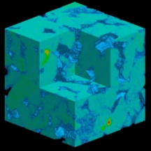
View of the structure of a Berea sandstone. The lighter colored portions represent solid material and the darkest regions are the pore spaces. Pore-scale calculations determine the flow rate through the experimentally determined microstructures. Courtesy of Keith Jones. |
 |
Geotimes Home | AGI Home | Information Services | Geoscience Education | Public Policy | Programs | Publications | Careers |